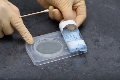
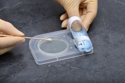
Product Principle:
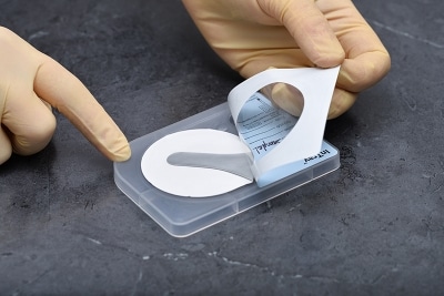
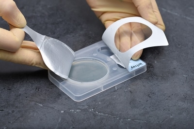
Simple Convenience:
Time Result:
Accurate Detection:
| Test Strain | ATCC # | Result |
|---|---|---|
| T. mentagrophytes | 9533 | Growth |
| T. rubrum | 28188 | Growth |
| M. gypseum | 14683 | Growth |
| A. brasiliensis | 16404 | Inhibited |
| S. aureus | 25923 | Inhibited |
| E. coli | 25922 | Inhibited |
| C. albicans | 60193 | Inhibited |
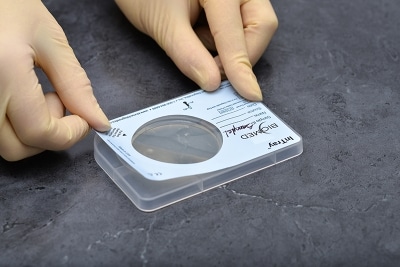
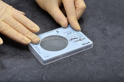
Storage:
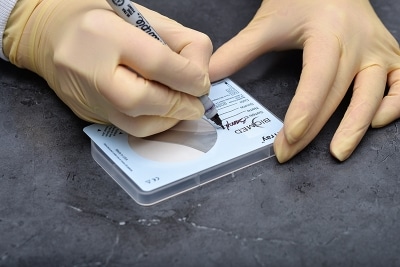
Specimen:
Trichophyton mentagrophytes | T. rubrum | T. tonsurans | Microsporum gypseum
Comparative study of different microscopic techniques and culture media for the isolation of dermatophytes.
Singh S, Beena P M.
Indian J Med Microbiol [serial online] 2003 [cited 2019 May 9 ];21:21-24
Diagnostic microbiology in veterinary dermatology: Present and Future
Luca Guardabassi, Peter Damborg†, Ivonne Stamm, Peter A. Kopp, Els M. Broens, and Pierre-Louis Toutain, the ESCMID Study Group for Veterinary Microbiology
Vet Dermatol 2017; 28: 146–e30
Principles and Practices of Veterinary Technology
Margi Sirois
Elsevier Health Sciences, Jul 19, 2016
Presentations
⚠ WARNING: This product can expose you to chemicals including Cycloheximide, which is known to the State of California to cause birth defects or other reproductive harm. For more information go to P65Warnings.ca.gov.
This product is for Veterinary Use Only.